Design Aspects of the Coloring Pages

Coloring page for plant and animal cells – The design of these coloring pages aims to be both engaging and educational, fostering a deeper understanding of plant and animal cells through visual representation and interactive learning. The use of clear labeling and comparative layouts will aid comprehension, making the complex structures of cells accessible to a wider audience. Coloring, a familiar and enjoyable activity, serves as a powerful tool for memorization and retention of key cellular components.
So you’re coloring plant and animal cells, huh? Feeling a little… underwhelmed? Spice things up! Learn to animate your cellular masterpieces with this awesome clip studio paint animation coloring tutorial , then go back to your coloring page and impress everyone with your newfound, hyper-realistic, possibly sentient, cell drawings. Who needs a microscope when you’ve got Clip Studio Paint?
The following sections detail the design specifications for each coloring page, focusing on clarity, accuracy, and visual appeal.
Plant Cell Coloring Page Design
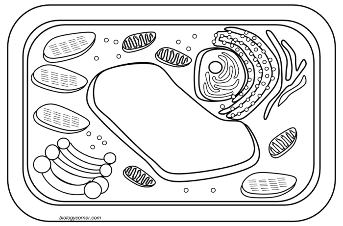
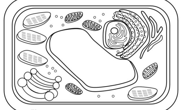
This coloring page features a detailed illustration of a typical plant cell. The design utilizes a two-column responsive layout for optimal viewing across different devices. The image column presents a visually appealing depiction of the cell, highlighting its various organelles. The second column provides clear and concise labels corresponding to the illustrated organelles.
|
Imagine a large, central, oval-shaped structure (the vacuole) dominating the cell’s interior. Surrounding this are numerous smaller, oval or bean-shaped chloroplasts, scattered throughout the cytoplasm. A clearly defined cell wall encases the entire cell, giving it a rigid, rectangular shape. Within the cytoplasm, a smaller, darker oval, the nucleus, is prominently displayed. Delicate, thin lines represent the endoplasmic reticulum, weaving throughout the cell. Finally, smaller, dot-like structures represent ribosomes, distributed near the endoplasmic reticulum. The entire cell is filled with a light green tint to represent the chlorophyll within the chloroplasts. |
|
Animal Cell Coloring Page Design
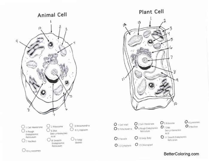
This coloring page showcases an animal cell, employing a three-column layout. The first column displays the illustration of the animal cell, the second provides labels for each organelle, and the third offers a brief description of its function. This multi-faceted approach ensures comprehensive learning.
|
The illustration shows a roughly circular cell, lacking a rigid outer wall. The nucleus is centrally located, but smaller than the plant cell’s. Mitochondria are depicted as numerous, bean-shaped structures scattered throughout the cytoplasm. The endoplasmic reticulum is represented by a network of thin lines, and ribosomes appear as small dots clustered near the ER. The Golgi apparatus is depicted as a stack of flattened sacs, near the nucleus. Lysosomes are small, circular organelles, and the cell membrane forms the outer boundary. |
|
|
Comparative Coloring Page: Plant and Animal Cells
This coloring page presents a side-by-side comparison of plant and animal cells, facilitating a direct understanding of their similarities and differences. The visual juxtaposition, combined with a concise list of key distinctions, enhances learning effectiveness.
The page features two illustrations, one of a plant cell and one of an animal cell, placed side-by-side. Key differences are highlighted through color-coding and labeling.
- Plant cells have a rigid cell wall; animal cells do not.
- Plant cells typically contain a large central vacuole; animal cells have smaller vacuoles, or none.
- Plant cells contain chloroplasts for photosynthesis; animal cells do not.
- Plant cells usually have a more rectangular shape due to the cell wall; animal cells are more variable in shape.
Illustrations and Descriptions
The following descriptions aim to paint a vivid picture of the cellular components, emphasizing their roles within the intricate machinery of plant and animal cells. Imagine these descriptions accompanying detailed illustrations, bringing the microscopic world into sharp, vibrant focus. The differences, subtle yet significant, between these two cell types will become clear as we delve into the specifics.
Chloroplast Structure and Function
Chloroplasts are the powerhouses of plant cells, responsible for photosynthesis. Imagine a vibrant green oval, filled with stacks of thylakoids – think of them as tiny, flattened sacs resembling a stack of pancakes. These thylakoids are embedded within a stroma, a gel-like substance. The chloroplast’s double membrane encloses this entire structure. Within the thylakoids, chlorophyll, the green pigment, captures light energy, initiating the process that converts carbon dioxide and water into glucose (sugar), the cell’s primary energy source, and oxygen, a byproduct released into the atmosphere.
The stroma plays a crucial role in the later stages of photosynthesis, where the captured light energy is used to convert carbon dioxide into glucose.
Mitochondrion Structure and Function
Mitochondria, often referred to as the “powerhouses” of both plant and animal cells, are oval-shaped organelles with a double membrane. The inner membrane is folded into cristae, creating a large surface area for cellular respiration. Picture these folds as intricate, shelf-like structures within the mitochondrion. Cellular respiration is the process where glucose is broken down to release energy in the form of ATP (adenosine triphosphate), the cell’s primary energy currency.
This energy fuels all cellular activities, from muscle contraction to protein synthesis. The space within the inner membrane, called the matrix, contains enzymes essential for this energy-generating process.
Nucleus Structure and Function, Coloring page for plant and animal cells
The nucleus, the cell’s control center, is a large, round organelle enclosed by a double membrane called the nuclear envelope. Imagine a sphere containing a dense, granular material. This material is chromatin, a complex of DNA and proteins. DNA, the cell’s genetic material, contains the instructions for building and maintaining the cell. The nucleolus, a smaller, dense region within the nucleus, is the site of ribosome production.
The nuclear envelope is punctuated by nuclear pores, which regulate the passage of molecules between the nucleus and the cytoplasm.
Cell Wall Structure and Function
The cell wall, a rigid outer layer found only in plant cells, provides structural support and protection. Imagine a strong, inflexible box surrounding the cell. It is primarily composed of cellulose, a complex carbohydrate that forms a tough, protective barrier. The cell wall maintains the cell’s shape, prevents excessive water uptake, and protects it from mechanical damage. It’s a key distinguishing feature separating plant cells from animal cells.
Endoplasmic Reticulum, Golgi Apparatus, Ribosomes, and Vacuoles
The endoplasmic reticulum (ER) is a network of interconnected membranes extending throughout the cytoplasm. The rough ER, studded with ribosomes, is involved in protein synthesis and modification. The smooth ER synthesizes lipids and detoxifies harmful substances. The Golgi apparatus, a stack of flattened sacs, modifies, sorts, and packages proteins and lipids for transport within or outside the cell.
Ribosomes, tiny structures found on the rough ER and free in the cytoplasm, are the sites of protein synthesis. Vacuoles are membrane-bound sacs that store water, nutrients, and waste products. In plant cells, a large central vacuole often occupies a significant portion of the cell’s volume, contributing to turgor pressure and maintaining cell shape.
Size and Shape Differences Between Plant and Animal Cells
Plant cells are typically larger and rectangular or cube-shaped due to the presence of the cell wall, which gives them a defined structure. Animal cells, lacking a cell wall, are generally smaller and more varied in shape, often round or irregular. Imagine comparing a neatly arranged stack of bricks (plant cells) to a collection of differently sized and shaped pebbles (animal cells).
The presence of a large central vacuole in plant cells also contributes to their larger size and distinct shape compared to animal cells.
Educational Activities and Extensions: Coloring Page For Plant And Animal Cells

The coloring pages, while seemingly simple, offer a potent springboard for deeper engagement with the intricacies of cell biology. They are not merely a passive exercise; rather, they serve as a visual scaffold upon which more complex understanding can be built. The activities described below aim to transform the coloring experience from a solitary activity into a dynamic learning journey.The coloring pages provide a foundation for several engaging educational activities, moving beyond simple identification to encompass a more nuanced understanding of cell structure and function.
These activities cater to diverse learning styles, fostering both individual exploration and collaborative learning.
Cell Organelle Identification Activity
This activity utilizes the completed coloring pages as a basis for a knowledge check. Students will be presented with a list of cell organelles (e.g., nucleus, mitochondria, chloroplast, cell wall, cell membrane) and asked to identify their location on their colored diagrams. This assessment can be administered individually or in small groups, promoting peer learning and discussion. A simple scoring system could be implemented, rewarding correct identifications.
For instance, a point system could be applied where each correctly identified organelle earns a point, and the total score reflects overall understanding. A visual aid, perhaps a separate key or labeled diagram, could be provided for those requiring additional support.
Lesson Plan Incorporating Coloring Pages and Quiz
A comprehensive lesson plan might begin with an introductory discussion on the fundamental differences between plant and animal cells. The coloring pages would then be distributed, allowing students to actively engage with the visual representation of this material. The coloring activity should be followed by a guided discussion, encouraging students to compare and contrast their completed diagrams. This discussion would address the functions of key organelles, emphasizing their significance within the larger context of cellular processes.
Finally, a short quiz could be administered to assess comprehension. This quiz could consist of multiple-choice questions, short-answer questions, or a combination of both. Example questions might include: “What is the function of the mitochondria?”, “What is the primary difference between the cell walls of plant and animal cells?”, or “Name three organelles found in both plant and animal cells.” The quiz questions would directly relate to the information presented in the coloring pages and the preceding discussion.
Questions Focusing on Plant and Animal Cell Differences and Similarities
Following the completion of the coloring pages, students should be prompted to reflect on their work by answering a series of questions designed to solidify their understanding of the differences and similarities between plant and animal cells. These questions should encourage critical thinking and comparison. For example: “Describe the differences in size and shape between a typical plant cell and a typical animal cell, as depicted in your coloring pages.” “Identify at least three organelles present in both plant and animal cells, and explain their shared function.” “Explain the role of the cell wall in plant cells, and why animal cells lack this structure.” “How do the vacuoles in plant and animal cells differ in size and function?” These questions are designed to facilitate deeper understanding beyond simple identification.
Students should be encouraged to use their completed coloring pages as a visual reference to support their answers.
Accessibility and Inclusivity
Creating coloring pages that are both engaging and accessible requires a thoughtful approach, considering the diverse needs and learning styles of our students. The aim is to ensure that every child, regardless of their abilities or background, can participate fully and benefit from the educational experience. This extends beyond simply providing a visually appealing image; it involves proactive design choices that promote inclusivity and cater to a wide range of learners.These coloring pages, designed to explore the intricacies of plant and animal cells, should be accessible to all students, regardless of visual acuity.
Tactile elements and alternative formats are crucial for students with visual impairments. Similarly, adapting the design to accommodate different learning styles—visual, auditory, kinesthetic—will ensure that every student can connect with the material in a meaningful way. The ultimate goal is to create a truly inclusive learning experience that celebrates diversity and fosters a sense of belonging for every participant.
Tactile Adaptations for Visually Impaired Students
Providing alternative formats for students with visual impairments is paramount. Raised-line drawings, created using materials like thick paper or cardboard, can allow students to trace the cell structures and internal components. These tactile representations offer a tangible way to understand the spatial relationships within the cells. Embossing techniques, or even simple, strategically placed glue dots to represent organelles, can also enhance the tactile experience.
Furthermore, accompanying audio descriptions that detail the cell structures and their functions can complement the tactile experience, providing a multi-sensory learning opportunity. Consider including braille labels for key organelles, further enriching the learning experience.
Adapting for Diverse Learning Styles
The coloring pages should be designed to cater to various learning styles. For visual learners, the illustrations themselves are key. Using clear, bold lines and vibrant, contrasting colors will enhance visibility and comprehension. For auditory learners, providing an audio component that describes the cell structures and functions will reinforce the visual information. This could include a narrated PowerPoint presentation or a short audio recording.
Kinesthetic learners benefit from hands-on activities. This could involve building 3D models of cells using clay or other materials, further solidifying their understanding of the spatial arrangements of organelles. Providing a range of activities will ensure that all students can engage with the material in a way that suits their learning preferences.
Promoting Inclusivity and Representation
To promote inclusivity, the coloring pages should avoid stereotypes and depict a diverse range of individuals engaging with the scientific content. The illustrations could show children of different ethnicities, genders, and abilities actively exploring plant and animal cells. This visual representation fosters a sense of belonging and encourages participation from all students. The language used in any accompanying materials should also be inclusive and accessible, avoiding jargon and employing clear, simple language.
The goal is to create a welcoming and engaging learning environment where every student feels valued and represented.

